Histological Techniques - Microtomy
- Microtomy - Paraffin/resin
- Microtome Section and stain
- Microtome Past
- Microtome Types
The procedure of microtomy takes place once the tissue has been hardened into a tissue block
Microtomes, as shown below, are used for production of thin tissue sections, varying between 1-10 microns, although the more normal range in a pathology laboratory is between 3-6 microns
Paraffin Routine Block. Paraffin Sidein Block. Resin block.
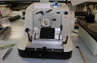

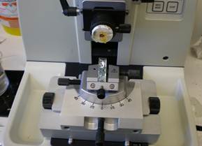
These microtomes are used in the routine lab for sectioning of paraffin embedded blocks, while in a resin histology lab, they are used for cutting tissue in resin eg bone marrows embedded in resin. Having the correct slant and tilt is especially important for resin sections as they are often not flat on the chuck, unlike paraffin blocks, which are normally flat on the cassette when embedded.
The reason for these microtomes being so commonly used in the laboratory and histological laboratories in general, is that these microtomes are able to produce truly flat sections, unlike rocking microtomes for example which often produce concave sections from the tissue.
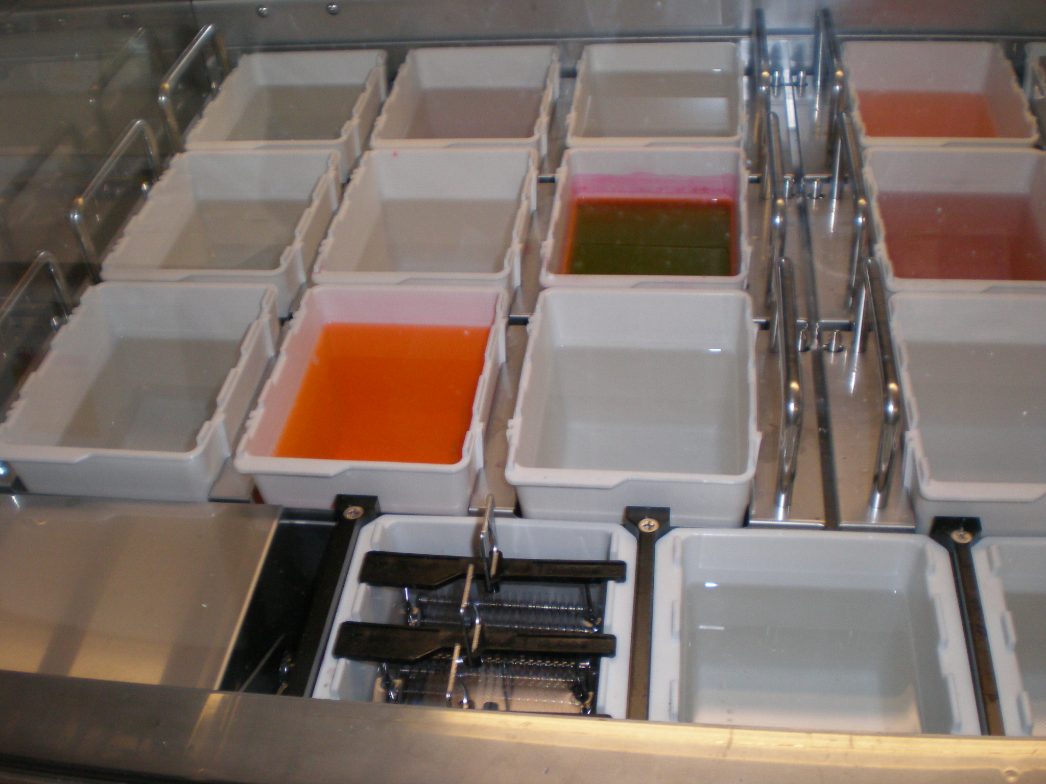
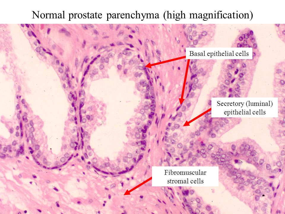
The section taken onto a slide then it is oven dried for around 20 - 30 minutes. Once dry the section is then stained, routinely with an H&E stain. In our lab we use a staining machine shown below, to produce an H&E stain, which is similar to the prostate case on the right side.
Microtomes are a sectioning instrument that is used for the sectioning of thin slices of material. Microtomes are an important device in microscopy as well. Microtomes can use a variety of materials for cutting whether it is steel, glass, or diamond blades depending upon the specimen being sliced and the desired thickness of the sections being cut. Steel blades are used to prepare sections of animal or plant tissues for light microscopy or routine histology. While Glass knives are used to slice sections for light microscopy and to slice very thin sections from tissue embedded in resin. Industrial grade diamond knives are used to slice hard materials such as bone, teeth and plant matter for both light microscopy very hard resin embedded tissue and for electron microscopy.
Here is a list of Historical Microtomes below
| Microtome Types and Functions | ||||||||||||||
|---|---|---|---|---|---|---|---|---|---|---|---|---|---|---|
| Type of Microtome | Functions | |||||||||||||
| Rotary Microtome | Most common microtome. Used for sectioning of Paraffin embedded blocks. Also can be used for Frozen sections in cryostat and also for resin embeded cases, for example Renal biopsies LR white resin embedded tissue and Bone marrows embeddded in methyl methacrylate (MMA). Sectioning occurs by movement of microtome head containing block across blade. | |||||||||||||
| Base Sledge Microtome | Heavy duty microtome that is able to cut celloidin embedded tissue from brain, gelatine embedded whole organs using Gough wentworth method and for sectioning undecalcified teeth and bone embedded in resin. | |||||||||||||
| Rocking microtome | Small microtome that has 2 rocking arms one to cut sections other to feed through tissue block. Limited to sectioning small soft blocks as it uses spring action to cut. | |||||||||||||
| Sliding Microtome | Unusual design microtome with blade moving over block, rather than block moving. Good for celloidin sectioning, although can produce good paraffin sections. | |||||||||||||
| Ultramicrotome | Microtome used mainly for Electron microscopy, for example for cutting Epon resin embedded sections. | |||||||||||||
| Hand Microtome | Very early microtome, with scalpel blade placed above micrometer screw. It is not useful in histology main use for botanical specimens. | |||||||||||||