Special Stains - Congo Red
- Amyloid stain
- Congo Red History
- Polarising Microscopy for Amyloid
- Congo Red Method
The main special stain used for amyloid detection is Congo Red. The reason for this is that the Congo red dye used in the stain is highly selective for amyloid deposits, which can lead to amyloidosis and also alzheimers.
This attachment is achieved by the non-polar hydrogen bonds formed between the Congo red and amyloid deposit.
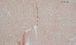
Under light microscopy the nuclei are stained up by haematoxylin blue, while the Amyloid, Elastic Fibres, Eosinophilic Granules and Keratin are stained up red, as shown below.
Photo - Congo Red under light microscopy
The first use of Congo red came in 1883 by an accident discovery by Paul Bottiger in Elberfeld, Germany. Originally intended of textiles, this use was abandoned but its use in histology due to its ability to detect amyloid has continued. This stain is considered the most accurate and efficient way for amyloid detection.
However to get an accurate detection of amyloid from the other potential background molecules, the section requires visualisation under a polarising microscope as shown below.
Photo 2 – Congo red under polarising microscope X10
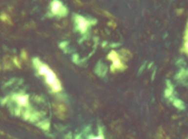
The apple green birefringence seen here, proves that it is infact amyloid that is been visualised rather than anything else. In the laboratory this can be used for diagnosis, for example of amyloidosis or alzheimers.
Below is the technique for performing this stain.
- Take sections to water. Sections cut at 6 -8 microns.
- Stain nuclei with alum haematoxylin, for 10 – 15 seconds
- Differentiate 1% Acid Alcohol
- Blue in Lithium Carbonate
- Wash in water
- Stain with 0.5% Congo Red in 50% alcohol for 10 minutes.
- Wash briefly in alcohol.
- Differentiate quickly in 0.2% Potassium Hydoxide in 70% alcohol
- Dehydrate, clear and mount
Results
Amyloid, Elastic Fibres, Eosinophilic Granules, Keratin |
Orange Red |
Nuclei |
Blue |
Amyloid – with polarised light |
Apple green (Birefringence) |
Photo - x10 Congo Red staining