
Special Stains - Auramine Rhodamine
- Bacteria - Auramine Rhodamine
- Auramine Rhodamine Technique
- Auramine Rhodamine History
Auramine rhodamine is used as a confirmation of the bacili, Following staining in the auramine rhodamine solution, the sections are differentiated in either 0.5% hydrochloric acid in alcohol for tubercle bacilli OR 0.5% aqueous hydrochloric acid for leprosy bacilli. The reason being that the leprosy bacilli is less acid and alcohol fast than the tubercle bacillus. Background tissue fluorescence is masked by the potassium permanganate solution. This technique employs the use of a fluorescent microscope for visualisation of the bacteria.
Below is the technique for performing the Auramine Rhodamine stain.
- Take sections to water
- Filter the auramine-rhodamine solution into a coplin jar.
- Preheat solution to 60º C (oven/waterbath)
- Place sections into the auramine rhodamine solution coplin jar for 10 Mins
- Wash in water for 2 mins
- Differentiate in 1% Hydrochloric Acid in Alcohol (tubercle Bacillifor 2-3 Mins OR 1% Hydrochloric Acid in Water (Leprosy Bacilli) 2-3 mins
- Wash in water
- Treat with 0.5% potassium permanganate for 1 Min.
- Wash in water
- Dehydrate, clear and mount
Results
Tubercle or Leprosy bacilli |
Golden Brown/Yellow |
Background |
Dark Green |
Photo - x10 Auramine Rhodamine IF fluorescent section
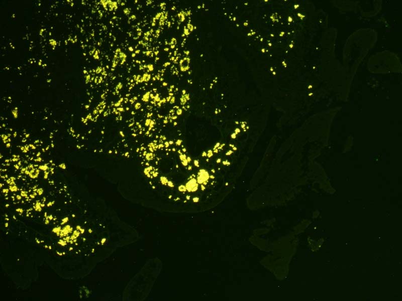
The auramine-rhodamine stain (AR), also known as the Truant auramine-rhodamine stain, is a histological technique used to visualize acid-fast bacilli using fluorescence microscopy, notably species in the Mycobacterium genus. Acid-fast organisms display a reddish-yellow fluorescence. Although the auramine-rhodamine stain is not as specific for acid-fast organisms (i.e. Mycobacterium tuberculosis or Nocardia) as the Ziehl-Neelsen stain, it is more affordable and more sensitive, therefore it is often utilized as a screening tool.
AR stain is a mixture of auramine O and rhodamine B, however care must be taken as this is a carcinogen.